Hair loss rarely begins with a clear label. It may appear as widening part lines, broken hairs, shiny patches, or sudden shedding in the shower. The American Academy of Dermatology reports that hereditary hair loss affects more than 80 million people in the United States. Meanwhile, the International Society of Hair Restoration Surgery’s 2022 Practice Census confirms continued global demand for hair-loss assessment and treatment. These figures show why earlier, more careful identification matters.
This guide explores how to identify hair loss types with a scalp camera, including androgenetic alopecia, alopecia areata, telogen effluvium, traction alopecia, and inflammatory scalp disorders. A scalp camera can reveal miniaturized hairs, follicular openings, scaling, redness, and uneven density. Dr. Antonella Tosti, a leading dermatologist and trichoscopy researcher, states: “Trichoscopy is a non-invasive technique that allows the diagnosis of hair and scalp disorders.” Her work supports camera-assisted observation as a valuable clinical tool, not a replacement for medical judgment.
The details can be surprisingly small. One follicle may hold three thin hairs. Another may show yellow dots or empty openings. Lighting, focus, hair products, and camera pressure can change the image. That part is easy to underestimate. A single photograph cannot confirm a diagnosis, and some patterns overlap. This article therefore treats scalp-camera findings as evidence, not certainty. Use consistent magnification, compare matched scalp areas, and record changes over time. When pain, scarring, rapid shedding, or inflammation appears, a qualified dermatologist should review the images. Some interpretations may remain imperfect, but careful observation is still better than guessing.
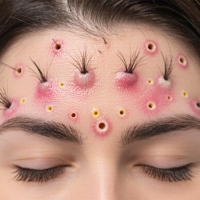
Hair loss does not look the same under a scalp camera. And that difference matters.
A close image can show thinner hairs beside normal strands, suggesting androgenetic hair loss. It may reveal round, smooth patches linked with alopecia areata. Broken hairs, uneven lengths, or scalp irritation can point toward traction or trichotillomania. Diffuse shedding may fit telogen effluvium, while sudden loss after medical treatment can suggest anagen effluvium. Other patterns include tinea capitis, scarring alopecia, loose anagen syndrome, and inherited hair-shaft disorders. The camera does not replace a medical examination. It makes small clues easier to compare.
Look for details, not dramatic pictures. A healthy follicle opening usually appears clear, while yellow dots, black dots, redness, scale, or absent openings may require professional review. A camera can also record density across the hairline, crown, and parting. Repeating images under similar lighting helps track change over time. Hair fibers can look thicker after washing, so timing affects interpretation.
The image can mislead.
Oil may resemble scaling. Bright light may exaggerate redness. One photograph cannot explain shedding caused by hormones, stress, nutrition, infection, or medication. Reliable identification combines camera findings with the shedding timeline, symptoms, family history, and, when needed, scalp examination or laboratory testing. I would not label a hair-loss type from one close-up alone. That limitation is easy to forget, especially when the image looks convincing.
| No. | Hair Loss Type | Typical Pattern or Onset | What a Scalp Camera May Show | Useful Distinguishing Clues | Important Limitation or Next Step |
|---|---|---|---|---|---|
| 1 | Androgenetic Alopecia | Gradual thinning; usually a receding hairline and crown loss in men, or widening of the central part in women. | Different hair-shaft diameters in the same area, increased fine or miniaturized hairs, reduced density, and more visible scalp. | Hair loss is usually patterned and slowly progressive. The frontal hairline, mid-scalp, or vertex may be more affected than the sides and back. | A camera can document miniaturization but cannot determine the cause alone. A clinician may correlate findings with history, examination, and family pattern. |
| 2 | Alopecia Areata | Often appears suddenly as one or more smooth, round or oval patches; severity can range from small patches to extensive loss. | Yellow dots, short regrowing hairs, tapered or exclamation-mark hairs, and broken hairs may be visible at patch margins. | Patchy loss with relatively smooth skin and no heavy scale is a common visual clue. Nails may also show changes in some people. | Similar appearances can occur in other conditions. Sudden patchy loss should be assessed by a qualified healthcare professional. |
| 3 | Telogen Effluvium | Diffuse shedding that often begins several weeks to a few months after illness, major stress, surgery, rapid weight loss, or hormonal change. | Reduced overall density with many short new hairs of similar length; the scalp may look fairly healthy without a distinct pattern. | Shedding is often widespread rather than localized. A clear trigger and recent increase in hair fall are important clues. | A camera cannot confirm the timing or trigger. Medical history and, when appropriate, laboratory evaluation are more informative. |
| 4 | Anagen Effluvium | Rapid, often extensive shedding during the growth phase of hair, commonly after certain medical treatments or significant toxic exposure. | Many broken or abruptly shortened hairs and a marked reduction in density across the scalp. | Hair loss usually develops quickly, often within weeks of a major exposure or treatment, rather than gradually over years. | The exposure and medical timeline are essential. Urgent medical guidance may be needed when rapid shedding follows treatment or serious illness. |
| 5 | Traction Alopecia | Hair loss caused by repeated pulling from tight hairstyles, extensions, braids, buns, or headwear that places prolonged tension on follicles. | Broken hairs, decreased density along tension points, short regrowth, and sometimes redness or follicular inflammation. | Loss commonly follows the hairline, temples, or areas under the greatest pull. Tenderness or bumps may occur early. | Continued traction can cause permanent follicle damage. Reducing tension promptly and seeking evaluation is advisable if loss persists. |
| 6 | Trichotillomania | Irregular, self-induced hair loss caused by repeated pulling or twisting, often producing uneven patches. | Hairs of many different lengths, broken shafts, tapered ends, and irregular patches rather than a consistent pattern. | The mixture of short and long hairs in the same area can be a clue. The person may not always recognize or report the pulling behavior. | A camera cannot establish behavior or emotional factors. Sensitive clinical assessment is important, and mental-health support may be helpful. |
| 7 | Tinea Capitis | A fungal scalp infection, more common in children, that may cause patchy hair loss, scaling, itching, or tenderness. | Broken hairs, black dots, scale, redness, crusting, or areas of inflammation may be visible. | Scaly patches, itch, swollen lymph nodes, or affected household contacts can support suspicion of infection. | A scalp camera cannot identify the fungus reliably. Medical examination and laboratory testing may be required; treatment generally needs prescription antifungal medicine. |
| 8 | Scarring Alopecia | Inflammatory disorders damage follicles and may cause permanent loss; progression can be gradual or active. | Reduced or absent follicular openings, shiny or scar-like skin, perifollicular scale, redness, pustules, or clumps of hairs may appear. | Loss of follicular openings and scalp inflammation are warning signs. Symptoms may include burning, pain, itching, or tenderness. | Early specialist assessment is important because established follicle destruction may be permanent. A biopsy may be needed for diagnosis. |
| 9 | Central Centrifugal Cicatricial Alopecia | Scarring hair loss that often begins near the crown and expands outward, most commonly affecting women of African descent. | Reduced follicular openings at the center of the scalp, perifollicular scale or redness, short broken hairs, and a widening central area. | Progression from the crown outward, scalp discomfort, or inflammation may help distinguish it from uncomplicated patterned thinning. | Camera images are useful for monitoring but not confirmation. Prompt dermatologic evaluation can help limit further permanent loss. |
| 10 | Hair Shaft Breakage | Hair appears thinner because shafts break from chemical processing, heat, friction, harsh grooming, or structural disorders. | Numerous broken hairs, split ends, frayed shafts, and uneven lengths with relatively preserved follicular openings. | Breakage may be concentrated where hair is exposed to heat, friction, or chemical treatments, while true shedding may be less prominent. | A camera can show shaft damage but cannot identify every underlying cause. Hair-care changes and clinical assessment may be needed if breakage is severe. |
It can show thinner hairs, broken strands, scaling, redness, and visible follicle openings. It shows patterns, not final diagnoses.
Mixed hair-shaft diameters and thinner crown strands may suggest hereditary pattern loss. Family history still matters. One image cannot confirm it.
It may show smooth, round patches with short exclamation-mark hairs. The surrounding skin can look normal. Professional assessment remains necessary.
Wash with a mild cleanser the day before, unless your clinician advises differently. Avoid oils, dry shampoo, sprays, fibers, and styling cream for 24 hours. Keep your scalp dry.
No. Arrive with your usual parting and avoid tight ponytails, braids, or extensions. These styles can irritate skin and alter hair direction temporarily.
Yes. Bright light may exaggerate redness, while oil can resemble scaling. Hair can also look thicker after washing. Images are not perfect.
Photograph the hairline, crown, temples, and parting under similar lighting. Keep the camera distance and hair condition consistent. Small differences can still confuse me.
Seek assessment for sudden loss, pain, pus, intense inflammation, or smooth shiny areas without follicle openings. These signs may indicate infection or scarring. Delay is not wise.
No. It cannot explain shedding from stress, hormones, nutrition, illness, or medication alone. Interpretation should include your timeline, symptoms, medicines, and family history. One close-up should not define you.
This guide explains how to identify hair loss types with a scalp camera by combining basic hair-loss knowledge with careful visual observation. It introduces why magnified scalp images can reveal details that are difficult to see with the naked eye, such as follicle density, hair shaft differences, scaling, redness, oil buildup, broken hairs, and areas of inflammation. It also covers how to prepare for a clearer examination, including using clean, dry hair and checking several scalp regions under consistent lighting.
The article reviews ten common hair loss patterns and the scalp clues that may help distinguish them, then presents a step-by-step camera method for comparing the hairline, crown, sides, and back of the scalp. Because similar signs can result from different conditions, camera findings should be treated as preliminary observations rather than a final diagnosis. Persistent shedding, sudden bald patches, pain, itching, or visible inflammation should be evaluated by a qualified healthcare professional.

 OneSe Beauty