Why is UV light used in skin analyzers? The answer begins with visibility. Standard lighting shows surface color, texture, and visible redness. UV illumination can reveal details beneath that appearance, including pigmentation patterns, accumulated sun exposure, and some porphyrin-related fluorescence.
“UV imaging can reveal skin changes that ordinary lighting cannot show,” explains dermatologist Dr. Zoe Diana Draelos. Her observation captures the practical value of this technology. A skin analyzer does not simply make a face look brighter. It creates contrast, helping professionals examine uneven tone and hidden patterns more consistently.
This guide explores seven reasons manufacturers and skincare specialists use UV light in skin analyzers. These reasons include detecting sub-surface pigmentation, observing oil-related fluorescence, identifying areas affected by environmental exposure, and supporting personalized skincare discussions. The images may look like blue, purple, or darkened facial zones on a monitor. Each color requires careful interpretation.
UV analysis is useful, but it is not a diagnosis. Results can vary with skin type, camera settings, room lighting, product residue, and operator experience. That limitation matters. A dramatic image can invite overconfidence, especially when a user expects certainty from technology.
Reliable systems need controlled lighting, calibrated sensors, hygienic procedures, and trained interpretation. Professionals should compare UV images with visible-light images and the person’s skin history. The technology supports observation. It does not replace clinical judgment. Some applications remain imperfect, and better standardization is still needed. Understanding these limits makes the seven tips more useful, practical, and trustworthy.
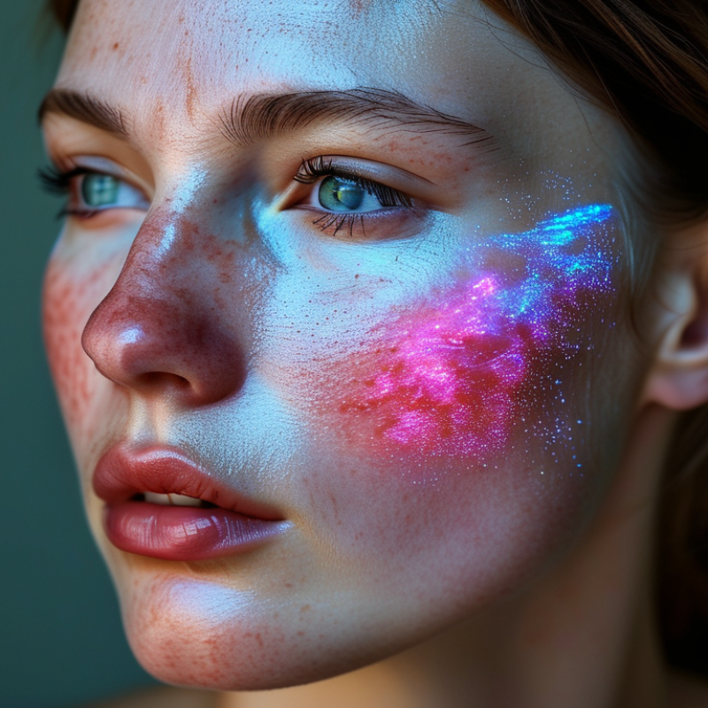
How UV Light Works in Skin Analysis
UV light helps skin analyzers reveal surface details that ordinary lighting can miss. Many systems use UVA wavelengths near 365 nanometres. Under this light, porphyrins may fluoresce around pores, while uneven pigmentation can appear darker. The U.S. Food and Drug Administration defines UVA as 320–400 nanometres. This range penetrates deeper than UVB, but analyzer images remain surface observations, not medical diagnoses. The World Health Organization reports two to three million non-melanoma skin cancers annually, showing why careful UV exposure education matters.
Tip 1: Use a calibrated device in a dim room. Ambient light can weaken contrast and create misleading shadows. Tip 2: Cleanse away makeup, sunscreen, and oil first. Residue may fluoresce and imitate skin concerns. Tip 3: Keep the camera distance consistent. Small changes can alter brightness and texture. Tip 4: Compare images over time, not from one session. Skin changes daily.
Tip 5: Record skin type and recent sun exposure. Melanin can affect UV contrast. Tip 6: Avoid interpreting every bright area as a problem. Fluorescence has several possible causes. Tip 7: Refer unusual, persistent, or changing findings to a qualified clinician. Analyzer results need professional context. A useful detail is often missed: device settings, room darkness, and operator technique can change the picture. That weakness deserves honest attention.
| No. | Analysis Focus | Typical UV Region | How UV Light Works | What the Analyzer May Reveal | Important Limitation |
|---|---|---|---|---|---|
| 1 | Surface oil and sebum mapping | Usually UVA, approximately 315–400 nm | Certain skin oils and residues can absorb or scatter UVA differently from surrounding skin, creating contrast in captured images. | Helps visualize areas that may appear oilier, especially across the forehead, nose, and chin. | Cosmetics, sunscreen, cleansing products, and recent washing can significantly alter the appearance. |
| 2 | Surface pigmentation contrast | UVA, commonly around 320–400 nm | Melanin absorbs ultraviolet radiation. Differences in melanin distribution can produce stronger image contrast than visible-light photography alone. | May make some sun-related pigmentation patterns and uneven tone easier to compare across the face. | The image is a visual aid, not a definitive measurement of melanin concentration or a medical diagnosis. |
| 3 | Early sun-exposure assessment | Primarily UVA, 315–400 nm | UVA penetrates more deeply than UVB and contributes to photoaging. UV-sensitive imaging can highlight contrast associated with cumulative exposure. | Supports educational discussions about sun protection and areas that may have accumulated visible or sub-visible changes. | UV imaging cannot determine the exact amount of past exposure or predict an individual’s future skin cancer risk. |
| 4 | Fluorescence-based observation | Often near-UVA, approximately 365 nm | Some substances emit visible fluorescence after absorbing UV energy. Skin surface materials and microbial by-products can therefore appear in contrasting colors. | Can assist with observing surface distribution, pore-area contrast, or features associated with follicular regions. | Fluorescence color and intensity are affected by lighting, camera settings, skin products, and individual skin chemistry. |
| 5 | Pore and follicle visibility | Usually UVA with specialized image processing | Differences in reflection, absorption, and fluorescence around follicular openings can increase local contrast in an image. | May help identify visually prominent pore areas and support before-and-after comparisons under controlled conditions. | Pore appearance also depends on focus, hydration, sebum, age, temperature, and image distance; it does not directly measure pore depth. |
| 6 | Standardized skin comparison | Controlled UVA illumination selected by the instrument | A fixed wavelength, illumination level, camera position, and exposure setting can reduce variability between repeated images. | Makes it easier to monitor changes in the same person over time when preparation and imaging conditions are consistent. | Results from different devices or sessions may not be directly comparable if their wavelengths, sensors, or software differ. |
| 7 | Enhanced visual education | Most commonly UVA; UVB is generally avoided for routine imaging | UV-based contrast can reveal information that is less obvious under ordinary visible light, helping explain skin-surface and pigmentation differences. | Provides an additional visual layer for skincare consultations, sun-protection education, and routine observation. | UV images should complement, not replace, clinical examination by a qualified healthcare professional when a lesion or persistent change is concerning. |
Safety note: Professional skin analyzers generally use controlled, low-intensity UVA illumination for brief imaging. Users should follow the device instructions, avoid staring directly into the light source, and use appropriate eye protection when required.
Near-UVA light, often around 365 nanometers, highlights pigments, oils, and surface compounds. Darker areas may show excess melanin or uneven sunscreen coverage. The image reveals clues, not certainty.
UV light interacts with substances that normal room lighting may not display clearly. It can reveal uneven pigmentation, rough patches, flakes, and clogged-looking areas. Mirrors miss details.
Orange-red spots may relate to porphyrins linked with certain surface bacteria. They can suggest congestion around pores. They do not prove acne or infection.
No. UV imaging cannot confirm cancer or measure every type of sun damage. A qualified medical professional must assess concerning changes with proper clinical testing.
Oil-rich areas may appear brighter under controlled UV illumination. The forehead, nose, and chin often show stronger signals than the cheeks. Temporary shine can confuse the result.
It may make rough, dull patches and tiny flakes easier to notice. Areas near the nose or mouth can appear uneven. The scan cannot directly measure every aspect of hydration.
Recent cleansing, heavy skincare, sweat, sunlight, cosmetics, and room lighting may change the image. Camera settings and calibration also matter. One scan can mislead.
Use clean skin and consistent lighting conditions. Record the room setup and repeat unusual scans when appropriate. Still, experience matters, and interpretation is not perfect.
This article explains why is UV light used in skin analyzers and how it provides information that ordinary lighting cannot reveal. UV light interacts with the skin’s surface and natural substances, creating differences in fluorescence and reflection. These patterns can help show hidden dryness, excess oil, accumulated dead skin, uneven pigmentation, and areas that may be more likely to develop acne. By making subtle changes visible, UV imaging supports a clearer understanding of skin condition and helps users choose more suitable skincare routines.
The article also highlights the importance of using UV analysis safely and accurately. Results should be interpreted as visual guidance rather than a medical diagnosis, since lighting conditions, skin tone, cosmetics, and device settings can affect the image. Proper calibration, clean skin, consistent testing conditions, and trained interpretation improve reliability. When used responsibly, UV skin analysis can make routine skin observation more informative while encouraging appropriate professional advice for persistent or concerning changes.

 OneSe Beauty